Purdue University Center for Cancer Research
Member Spotlight
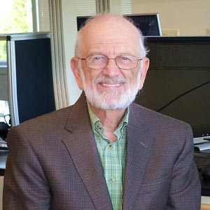
GRAHAM COOKS, PhD
Henry Bohn Hass Distinguished Professor – Analytical Chemistry
Director of the Aston Laboratory of Mass Spectrometry
Graham Cooks: on the forefront of brain cancer research
The innovative “DESI” mass spectrometer technique identifies tumor in tissue quickly and accurately
Cooks focuses on ways mass spectrometry can detect and diagnose cancerous tissue, particularly in brain cancers. He and his team are internationally recognized for devising a mass spectrometry tool called Desorption Electrospray Ionization, or DESI, which surgeons can use in the operating room to detect tumor margins – which is especially difficult for brain tumors.
The Walther Cancer Foundation provided critical funding for Cooks and his ideas — at a time when he had no federal funding for the research. Indeed, the Walther Foundation’s early funding for Cooks and DESI illustrates the foundation’s philosophy of focusing on “high risk, high reward” research.
The brain is different
In many kinds of cancers, there are well-established alternatives to surgery. With liver cancer, for example, it is possible to remove part of the organ; with prostate cancer, it is possible to remove the organ entirely.
In the brain, surgeons want to remove as little tissue as possible. In the operating room, it can be difficult to visualize, much less measure, the edges of the tumor that need to be removed.
Mass spectrometry presents a different approach for surgeons, one that involves making precise chemical measurements — the measured compounds are small molecules: lipids and metabolites. The technique leads to rigorous answers in the surgical suite.
In a matter of seconds, DESI provides the molecular information necessary for detecting tumor in small samples and residual tumor cells that otherwise may be left behind in the patient.
How it works
During surgery, the surgeon will biopsy the tumor’s margin. Using the DESI technique, the biopsied tissue is sprayed with a microscopic stream of charged solvent.
A software program, also designed by Cooks’ research team, matches the mass spectrometer profile of the current tissue sample against previously identified lipid patterns corresponding to different types and grades of cancer.
The DESI database includes the two most common types of brain tumors, gliomas and meningiomas, which account for 65% of all brain tumors and 80% of all malignant brain tumors, according to the American Brain Tumor Association.
Rapidly, DESI provides a color-coded image that reveals the nature and concentration of tumor cells. The surgeon can then see more clearly which brain tissue to remove — and which to leave intact — rendering the tumor removal procedure more exact — and safer.
The instrumentation is relatively small and inexpensive and could easily be installed in operating rooms to aid neurosurgeons.
Identifying aggressive mutations
In brain cancers, surgeons in particular look for mutations in the isocitrate dehydrogenase (IDH) enzyme. IDH enzymes are meant to help cells break down nutrients and generate energy. IDH mutations, however, lead to harmful alterations in a cell’s genetic programming and are a characteristic of particularly aggressive cancers.
In current practice, it is not possible to identify IDH mutations during surgery. Subsequent pathologic results may require another surgery for a patient who has been identified to have a cancer tumor with IDH mutations.
Another important use of DESI is that it allows surgeons to identify these IDH mutations with high sensitivity and specificity within minutes, immediately providing critical diagnostic, prognostic and predictive information.
Based on 10 years of clinical investigations, Cooks estimates that DESI-based surgeries can add two-to-three years to a brain-cancer patient’s life.
For Cooks, the next course of action for DESI is to continue with the above-described data collection and then prepare for clinical trials.